Pocket Atlas of Radiographic Positioning
(Sprache: Englisch)


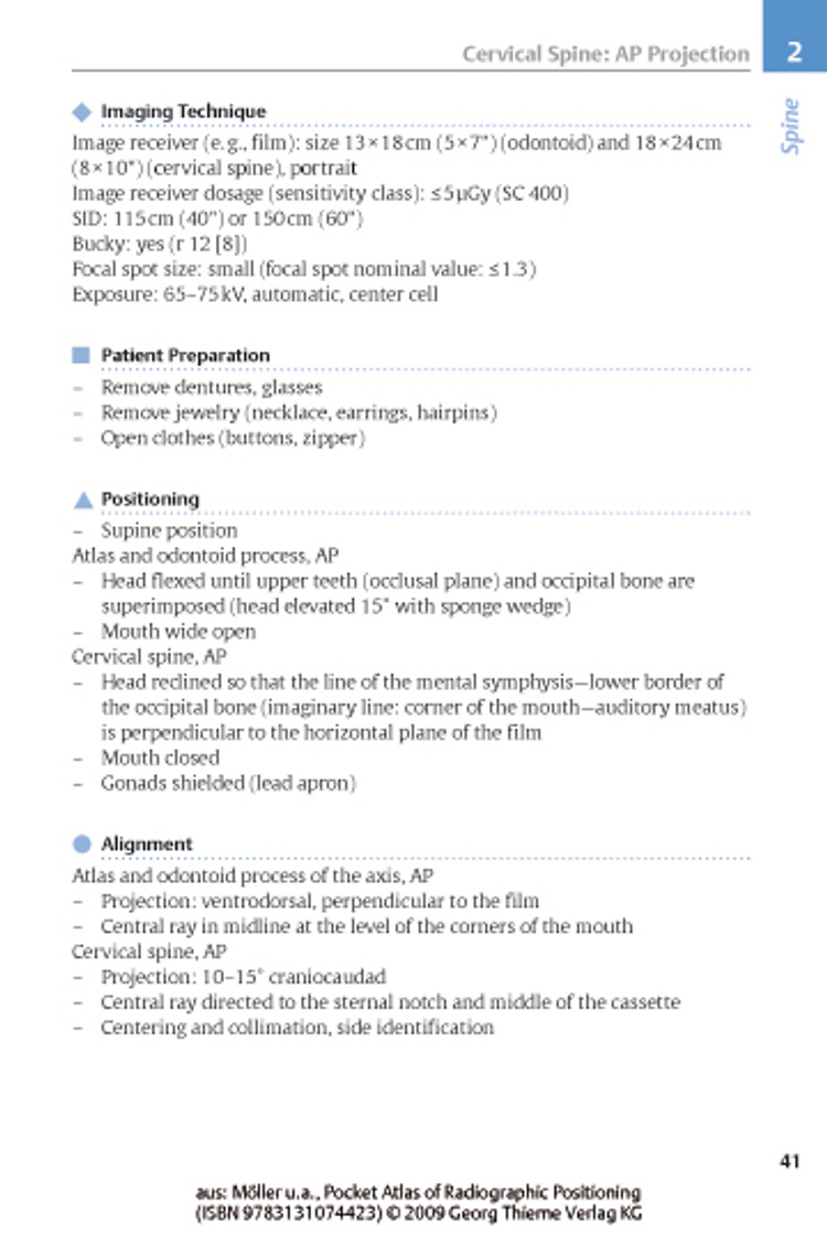


Flexibooks (clinical sciences) Accurate diagnosis depends on properly conducted radiographic imaging. Inappropriate positioning or exposure can lead to diagnoses being missed or incorrect. This handy atlas for radiologic technicians and physicians...
lieferbar
versandkostenfrei
Buch (Kartoniert)
52.50 €
- Lastschrift, Kreditkarte, Paypal, Rechnung
- Kostenlose Rücksendung
- Ratenzahlung möglich
Produktdetails
Produktinformationen zu „Pocket Atlas of Radiographic Positioning “
Klappentext zu „Pocket Atlas of Radiographic Positioning “
Flexibooks (clinical sciences) Accurate diagnosis depends on properly conducted radiographic imaging. Inappropriate positioning or exposure can lead to diagnoses being missed or incorrect. This handy atlas for radiologic technicians and physicians demonstrates not only how to position patients, but also how to ensure that the images obtained are optimal for diagnosis. The clear way in which the information is arranged for each type of study, including imaging technique and parameters, positioning, and, where appropriate, tips and tricks, makes the whole process much easier. Criteria for a good radiographic view are demonstrated on actual radiologic images. The updated and enlarged 2nd edition now includes all standard positioning and examination techniques for conventional radiology, CT and MR imaging. In the completely new chapter on computed tomography, the use of multidetector CT scanners is covered. Also new to this 2nd edition are the chapter on MR imaging and the much-expanded chapter on mammography. This Pocket Atlas of Radiographic Positioning is the ideal companion to Moeller and Reif's Pocket Atlas of Radiographic Anatomy and their three-volume Pocket Atlas of Cross-Sectional Anatomy.
Radiographic positioning- comprehensive and concise!
Now in its second edition, Pocket Atlas of Radiographic Positioning is a practical how-to guide that provides the detailed information you need to reproducibly obtain high-quality radiographic images for optimal evaluation and interpretation of normal, abnormal, and pathological anatomic findings. It shows positioning techniques for all standard examinations in conventional radiology, with and without contrast, as well as basic positioning for CT and MRI. For each type of study a double-page spread features an exemplary radiograph, positioning sketches, and helpful information on imaging technique and parameters, criteria for the best radiographic view, and patient preparation. Clearly organized to be used in day-to-day practice, the atlas serves as an ideal companion to Moeller and Reif's Pocket Atlas of Radiographic Anatomy and their three-volume Pocket Atlas of Sectional Anatomy.
Highlights of the second edition:
- New chapters on positioning for MRI and CT, including multislice CT
- A greatly expanded section on mammography
- Special features, including information on the advantages of a specific view, variations of positions, and practical tips and tricks
- Nearly 500 excellent radiographs and drawings demonstrating the relationship between correct patient positioning and effective diagnostic images Pocket Atlas of Radiographic Positioning, Second Edition is an excellent desk or pocket reference for radiologists, radiology residents, and for radiologic technologists and technicians.
Inhaltsverzeichnis zu „Pocket Atlas of Radiographic Positioning “
Skeletal Diagnosis:; - Skull; - Spine; - Upper Extremities; - Lower Extremities; Other Non-Contrast Diagnostic Studies incl. Mammography Contrast Examiniations:; - Gastrointestinal Tract; - Intravenous Examinations; - Angiographies; CT Examinations; MR Imaging.
Autoren-Porträt von Torsten B. Möller, Emil Reif
Emil Reif, MDDepartment of RadiologyMarienhaus Klinikum Saarlouis - DillingenDillingen/Saarlouis, Germany
Bibliographische Angaben
- Autoren: Torsten B. Möller , Emil Reif
- 2009, 2nd ed., 358 Seiten, 491 Abbildungen, Maße: 12,8 x 18,8 cm, Kartoniert (TB), Englisch
- Übersetzer: Horst N. Bertram, Michael Robertson
- Verlag: Thieme, Stuttgart
- ISBN-10: 3131074426
- ISBN-13: 9783131074423
Sprache:
Englisch
Rezension zu „Pocket Atlas of Radiographic Positioning “
Any pocket-sized handbook that provides in-depth coverage of multiple medical imaging modalities is a rare and remarkable thing. The second edition of "Pocket Atlas of Radiographic Positioning" is just such a book, containing nearly every modality, including conventional radiography, CT, MRI, angiography, and mammography...a valuable, easy-to-use desk or pocket reference for medical imaging professionals at every level.--ADVANCE for Imaging & Radiation Oncology"We suggest this publication not only to radiologists, radiology residents, radiologic technologists and technicians, but also to professionals and students working in nuclear medicine to acquire autonomous knowledge of instruments and information. European Journal of Nuclear Medicine and Molekular Imaging March 2010"This pocket book presents in a condensed but comprehensive manner the proper positioning for all radiographic procedures...This second edition has been enriched by new chapters on positioning in MRI and CT, including multislice CT, and by an expanded section on mammography...Each positioning is documented by clear drawings and by the related radiological image, except for the positions in CT and MR studies, which are presented by drawings, albeit very demonstrative...a solid companion to the three volumes Pocket Atlas of Sectional Anatomy, in which the same authors present, in a comprehensive and clear manner, the basic positioning and related radiographic images for CT and MR...will be of great value in the day-to-day practice of the radiologic technologists, but in particular it will be very useful to the Residents in Radiology.--Clinical ImagingA comprehensive pocket-sized guide of general radiography techniques, with lots of suggestions for different adaptations if the standard positions are not possible. With each technique there is a particularly good section Criteria for a Good Radiographic View which gives guidance on the optimum image, including the area or joint space that should be
... mehr
visu
... weniger
Kommentar zu "Pocket Atlas of Radiographic Positioning"
Schreiben Sie einen Kommentar zu "Pocket Atlas of Radiographic Positioning".
Kommentar verfassen